

Overview
The human brain can be organized into discrete neurocognitive systems, each handling aspects of cognition. Our long-term goal is to characterize how concepts are represented in these networks, and how those representations make contact with other neurocognitive and sensory networks.
A major research focus has been on phenomena related to naming objects, as this requires the successful integration of lexical-conceptual representations in the left-hemispheric language network with various object recognition networks (visual, auditory, olfactory), and with motor systems for speech production. Given this level of synaptic complexity, it is perhaps understandable that inability to name objects is one of the most common symptoms of brain injury in general, and is the most common symptom in acquired language disorders (aphasias). Difficulty in retrieving proper names is also the most frequent complaint associated with cognitive aging. We use online techniques including eye tracking and event-related potentials to examine information processing in real time as participants attempt to identify and match words and objects, in an attempt to better understand the neurocognitive mechanisms of naming.
Functional magnetic resonance imaging (fMRI) provides another way to characterize neurocognitive systems. Functional connectivity analysis can be used to locate key nodes, examine interactions between networks, and to examine how brain injury affects network coherence.
Some examples from each of these approaches are provided below.
Online Techniques
Eye tracking
Eye movements provide a window into the mind: if you know where someone is looking, you know much of what they are thinking at any given moment. In order to reveal the cognitive mechanisms of object naming, we examine eye movements as participants search for words or objects on the screen. Participants with neurodegenerative disorders of language, known as progressive aphasias, show distinct patterns of eye movements during these tasks. For example, individuals with atrophy in the left anterior temporal lobe (ATL) show a phenomena of “taxonomic blurring”, such that they repeatedly look back and forth between objects from the same semantic category (see videos below). These findings suggest that ATL is critical for the specific linkage between a visual object and its concrete noun label, with lesions in that area creating uncertainty and coordinate errors in object naming. Ongoing studies focus on the subtle differences in eye movements that typical adults show when recognizing words versus objects, and how those visual search patterns are altered by progressive aphasia and visual agnosia (impairment in recognizing objects).
Control Participant
![]() Neurocognitive Systems Laboratory - eye-tracking with control participant
Neurocognitive Systems Laboratory - eye-tracking with control participant
Participant with ATL lesion
![]() Neurocognitive Systems Laboratory - eye tracking with participant with ATL lesion
Neurocognitive Systems Laboratory - eye tracking with participant with ATL lesion
Event-related potentials
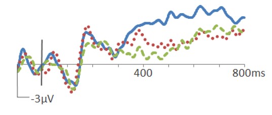
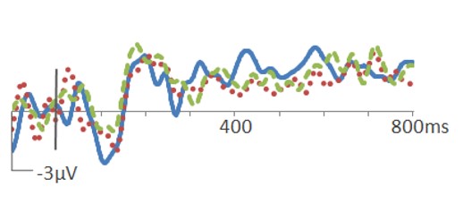
The averaged EEG response to stimuli, known as event-related potentials, are another online technique that has proven useful for the study of object naming. Naming failures are frequent in older adults and in individuals with language and object recognition disorders, and can be caused by any of a number of underlying neurocognitive mechanisms, including failures in object recognition, lexical access, or speech production. By indexing synaptic activity in real time as participants complete object-word matching tasks, event-related potentials can help reveal whether naming impairments have a perceptual, conceptual, or motoric basis, and can help localize this impairment to the relevant neurocognitive network. For example, naming impairments in participants with progressive aphasia are associated with reductions in N400 amplitude (see figure below), when judging words but not when judging pictures. Although these individuals report feeling that the name is on the “tip of their tongues”, these results suggest their naming impairments are based on a deeper lexical-conceptual mechanism in the left hemispheric language network. Current studies are examining perceptual and conceptual aspects of auditory word recognition in typical adults, and audiovisual linkage in progressive aphasia.
| ERPs to named items | ERPs to misnamed items |
|
|
|
Functional Connectivity
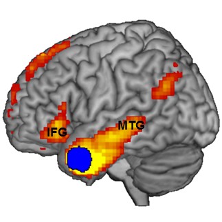
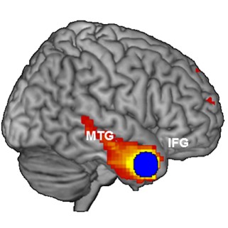
Functional magnetic resonance imaging (fMRI) reveals the hemodynamic byproducts of neural activity. By correlating spontaneous hemodynamic fluctuations across fMRI voxels, functional connectivity analysis reveals the extent to which brain regions are in communication with one another. In our laboratory we use functional connectivity to define the major nodes and boundaries of neurocognitive networks. For example, after placing “seeds” (blue in the figure below) in ATL, we showed that the lateral ATL in the left hemisphere is functionally connected with nodes of the language network such as Broca’s area (IFG), while ATL in the right hemisphere shows no such affiliations.
| Left Hemisphere | Right Hemisphere |
 |
 |
We are currently examining how functional connectivity in the language network is impacted by progressive aphasia, and how this relates to structural MRI. For example, do physiological changes precede morphological changes in the course of neurodegenerative disease? In order to better address these questions we have developed the Individually-Masked Group-Analyzed (IMGA) toolbox, which we are releasing as a freely available SPM extension. See the IMGA tab for details and downloads.